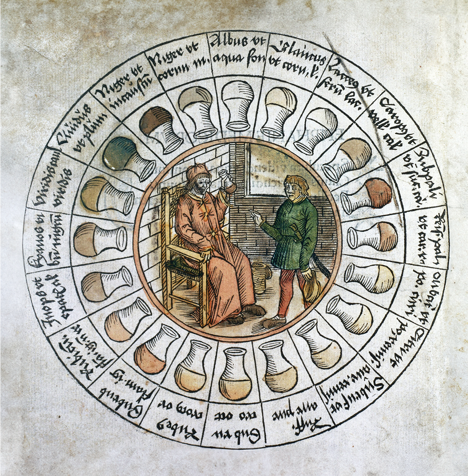
Figure 1. The “Urine Wheel” devised by Gilles de Corbeil.
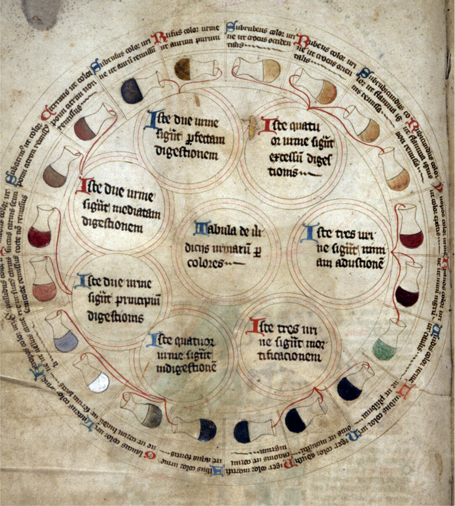
Figure 2. A medical ring diagram, comprising urine flasks from MS Savile 39, is dated to the period between ca. 1387 and 1400 (8).
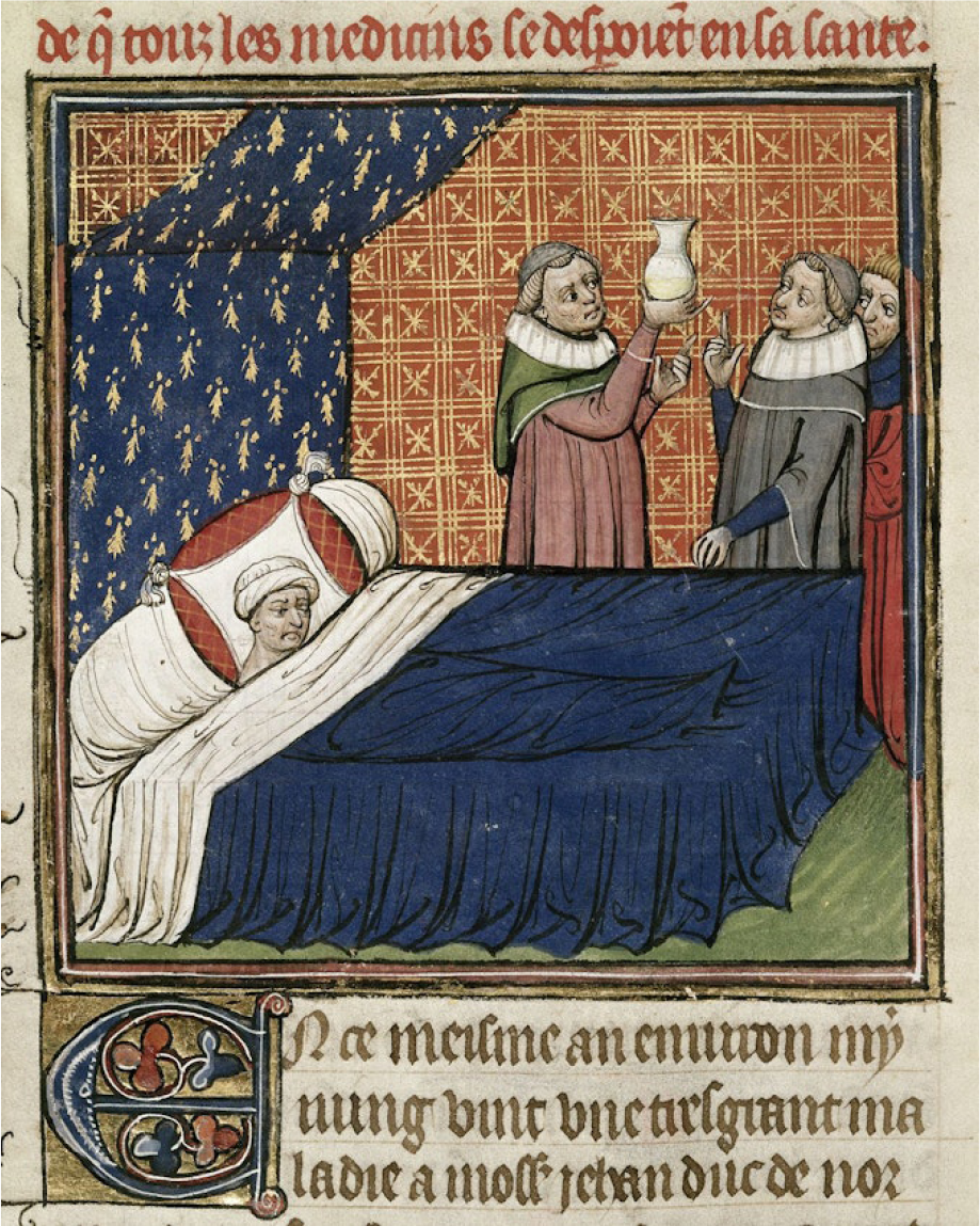
Figure 3. A detailed illustration of a miniature portraying the bedridden John, Duke of Normandy, and medical professionals examining his urine. The image was sourced from the Chroniques de France ou de St. Denis. The work was first published or produced at the end of the 14th century. (9)
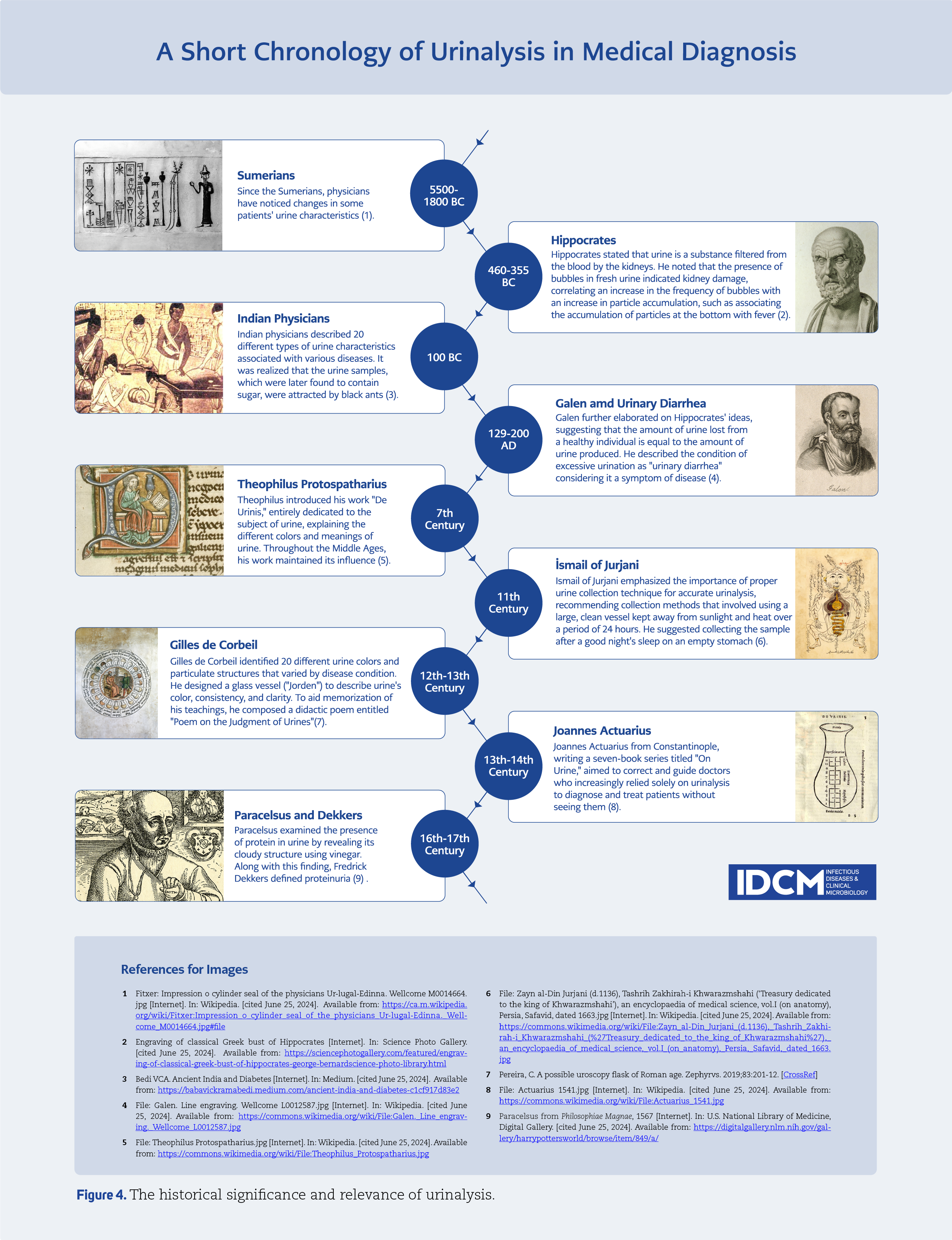
One of the indispensable abilities of a young medieval doctor was the capability to read urine colour, given that urine was regarded as a divine fluid and considered to be a window to the human body. The observation of urine enabled the detection of bodily changes, and it served as the first laboratory test for thousands of years. Similarly, the practice of uroscopy, which is currently referred to as urinalysis, has a long history, dating back to the 4th century BC. (1, 4) In Greek medicine, urine was observed, not for diagnostic, but rather for prognostic proposes. Hippocrates’ ( 460-370 BC) criteria for urine observation reportedly included factors such as colour, texture, sediment, odour and volume. Galen (129-216 AD ) developed these criteria and argued that body secretions could prove various types of internal imbalance A spectrum of colours and consistencies was employed to describe urine, as evidenced by medieval manuscripts. A colour description chart was created as a means of standardising the evaluation of urine by Gilles de Corbeil (1165–1213), and it described the “Urine Wheel” (Figure 1), of which colour spread is in Ulrich Pinder’s medical text, Epiphanie medicorum, printed in 1506 in Nuremberg, Germany (5). The Urine Wheel comprised 20 different conditions, each represented by a distinct colour and sediment. The vessels were shaped with a bladder-like bottom, allowing them to be held with one hand. Additionally, Gilles de Corbeil proposed a correlation between the various parts of the vessel and the corresponding parts of the body (1). The practice of urinalysis has been a significant clinical pathway for the treatment of medical conditions for thousands of years, with various forms having been employed. Since ancient times, the historical significance and relevance of urinalysis have been demonstrated (Figure 4), and it is still used today as an important diagnostic approach (1-3). It is imperative to evaluate urinalysis in conjunction with the patient’s clinical presentation, including age, gender and co-morbidities considerations (2, 3).
Current Use of Urinalysis in Clinical Practice
Currently, urinalysis is less important than it once was and is not typically considered a prerequisite for clinical diagnosis. However, in instances where the clinical presentation lacks sufficient specificity as a popular screening test, it may serve as an additional tool (1, 2, 6). It can be a useful guiding tool in diagnosing various conditions such as kidney stones, infections, diabetes, acute or chronic kidney injury, and metabolic disorders (2).
Pyuria, hemocytometry, and dipstick testing represent essential laboratory diagnostic modalities. Pyuria is commonly encountered in specimens from patients with urinary tract infections (UTIs), and its absence strongly suggests an alternate diagnosis. The presence of more than 10 cells per high-power field (hpf) on hemocytometry further supports the presence of pyuria.. The presence of leukocyte esterase on dipstick analysis is indicative of pyuria, while the conversion of nitrate to nitrite reflects the presence of Enterobacterales, offering a sensitivity of 75% and specificity of 82% (7). In cases, where the clinical history points towards a UTI, the absence of positive results from both tests does not definitively rule out infection. However, the urinalysis is still one of the most frequently ordered tests in our clinical practice.