Introduction
HIV infection remains a major global health concern with a considerable mortality rate, particularly in individuals with AIDS and related infections. Opportunistic infections, including tuberculosis (TB) and Kaposi’s sarcoma (KS), are common due to HIV-induced immunosuppression (1). Managing such co-infections and complications, including immune reconstitution inflammatory syndrome (IRIS), is critical for patient survival and health outcomes. In this case report, we present a patient with AIDS who developed KS-IRIS and disseminated TB.
Case
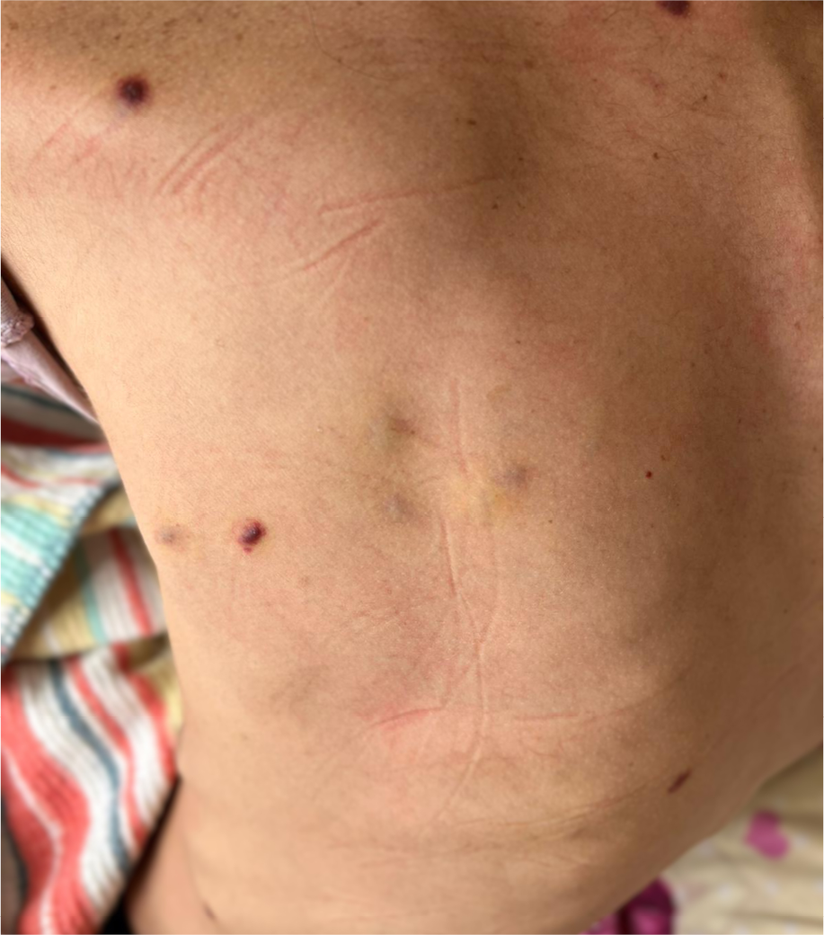
Figure 1. The rashes showing diffusely distributed, domeshaped nodules and livid plaques, with erythematous areas at the base.
A 25-year-old male case presented with a 20-day history of nausea, vomiting, headache, abdominal pain and a 6-kilogram weight loss. He also had a recent history of high fever. The patient had been diagnosed with HIV infection in 2018 but was not receiving regular antiretroviral therapy (ART). On the first day of admission, the patient was initiated on a regimen comprising tenofovir disoproxil fumarate-emtricitabine plus dolutegravir, which he was unable to adhere to consistently. The CD4 count was 7 cells/µL, and the plasma HIV-RNA viral load was over 400,000 copies/mL. Cranial magnetic resonance (MR) imaging showed a lesion 6.5 mm in diameter with peripheral contrast enhancement and surrounding vasogenic edema in the medial part of the left occipital lobe, consistent with the presence of an abscess. However, the lesion did not show any regression following 14 days of antibiotic treatment including meropenem 3x 2gram+ vancomycin 2×1 gram. Subsequent re-evaluation by a radiologist suggested a tuberculoma.
Cerebrospinal fluid (CSF) was clear in appearance, with no leukocytes or erythrocytes, and had normal glucose and protein levels. Gram staining revealed no leukocytes or microorganisms. CSF Culture and polymerase chain reaction (PCR) for tuberculosis (TB) were negative. Serum Toxoplasma gondii IgM/IgG, serum/CSF Cryptococcus neoformans antigen and CSF JC virus were all negative. CSF was sent out for analysis with multiplex PCR (herpes simplex virus 1 and 2, adenovirus, varicella zoster virus, enterovirus, cytomegalovirus, parechovirus, human herpes virus type 6, and Cryptococcus neoformans/gattii). All results were negative. In addition, F-18 FDG PET/BT revealed generalized lymphadenopathies (abdominal LAPs SUVmax= 7.42, mediastinal LAPs SUVmax:8.37, supraclavicular LAPs SUVmax:5.46), pleural effusion, terminal ileum/ileocecal wall thickening and vertebral osteomyelitis with the enhancement of L4 vertebrae. Bone marrow and ileocecal biopsy specimens were consistent with TB on histopathologic examination, and thetissues were also positive for acid-fast bacilli and TB PCR. A diagnosis of disseminated TB was made, and quadruple anti-TB drugs were initiated on the 14th day of ART. However, on day 30 of TB treatment, his general condition deteriorated, accompanied by the onset of fever, severe headache, and diplopia. Hemocultures were sterile, and extensive investigations were inconclusive. Control MR revealed an increase in the size of the lesion and the surrounding edema, attributed to IRIS, and systemic corticosteroid was initiated. Seven days later, multiple skin eruptions developed, first appearing on the upper limbs and then gradually spreading over the trunk. The lesions were multiple, diffusely distributed, dome-shaped nodules and livid plaques, 1 to 2 cm in size, with erythematous areas at the base (Fig. 1). Skin biopsy confirmed the presence of Kaposi’s-Sarcoma associated herpesvirus and was compatible with KS. The patient was diagnosed with unmasked KS-IRIS and admitted to the intensive care unit. The bone marrow culture yielded multi–drug resistant Mycobacterium tuberculosis complex that was sensitive to ethambutol but resistant to isoniazid, rifampicin, and streptomycin. Despite aggressive treatment, the patient succumbed to multiorgan failure in the hospital on day 85 of ART.
Discussion
We presented a case of disseminated TB with multiple organ involvement. Following the steroid treatment for IRIS, cutaneous lesions appeared on the extremities and trunk. Initial differential diagnoses for the skin eruptions included KS, bacillary angiomatosis, cutaneous TB, cutaneous lymphoma, and angiosarcoma. KS was finally confirmed by biopsy.
IRIS is characterized by an excessive inflammatory response upon immune reconstitution, a known complication of ART initiation. The incidence of IRIS is variable, with estimates ranging from less than 10% to over 50% in specific populations (2). Studies suggest that low CD4 counts and high HIV RNA levels at treatment initiation increase the risk of IRIS (3). Given that TB is one of the leading causes of death among people living with HIV, the initiation of prophylactic steroids in addition to anti-TB treatment in patients with a CD4 count below 100 cells/µL for IRIS prophylaxis could be a useful strategy. In this case, the patient’s critically low CD4 count and high HIV viral load likely contributed to the development of IRIS.
While the incidence and mortality rates of KS have decreased due to ART, it remains the most common malignancy in HIV-infected populations (4). The risk of KS is notably higher in patients with low CD4 counts and high HIV viremia, though it can occasionally occur in those well-controlled on ART (5). KS-IRIS is rare and can present as paradoxical IRIS (worsening of existing KS) or unmasked IRIS (emergence of new KS). The latter form, as observed in our patient, is associated with a high mortality rate (6, 7). The use of corticosteroids has been associated with the emergence and exacerbation of KS-IRIS (8).
In conclusion, this case underscores the complexity of managing AIDS patients with opportunistic infections. The use of corticosteroids can increase the risk of KS-IRIS and Kaposi’s sarcoma-related mortality, highlighting the need for cautious administration and close monitoring to detect KS lesions in such patients.